
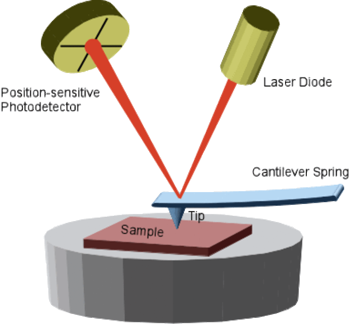
SEM picture of tip The Atomic Force Microscope (AFM) is one type of scanning probe microscopes, which is used to image surface structures (on a nm or even sub-nm scale scale) and to measure surface forces.
The standard AFM contains a microscopic tip (curvature radius of ~10-50nm) attached to a cantilever spring.
The underlying principle of AFM is the detection of the bending of this cantilever spring as a response to external forces. In the case of adhesive interaction between the tip and a surface, this forces are of the order 0.1 - 1 nN. To measure such small forces one must use not only very sensitive force-measuring springs but also very sensitive ways for measuring their bending.
In order to detect this bending, which is as small as 0.01 nm, a laser beam is focused on the back of the cantilever. From there the laser beam is reflected towards a position-sensitive photodetector. Depending on the cantilever deflection the position of the reflected beam changes. The photodetector converts this change in an electrical signal.
Direct Force Measurement
A way to get information on the surface forces wich are acting in a given system and to illustrate the functioning of an AFM, is to use the Force Mode. This is done by moving the tip towards and away from the surface. Recording photodetector signal as a function of z-piezo elongation yields a curve which can be interpreted as a force-vs.-distance-curve.
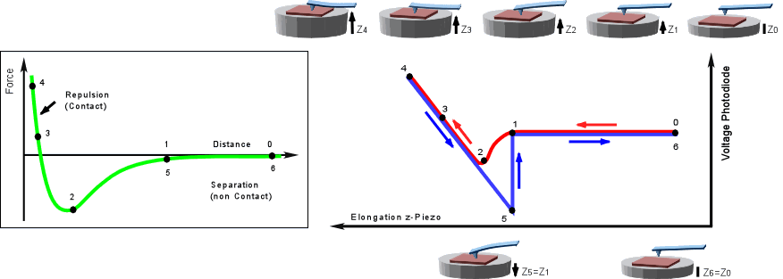
Asuming that the tip-surface interaction can be described by a Lennard-Jones-Potential one can observe a cycle as follows: (0) the tip is far away from the surface, and surface forces do not act. As the tip approaches (1) to (2), it enters the range of attractive surface forces and is deflected downwards. (2) to (4): the tip is in contact with the surface and now exerts pressure while it is deflected upwards. At (4) the tip retraction started, but adhesion forces may keep the tip attached to the surface until the spring force exerted by the cantilever can overcome adhesion (5). Then the tip snaps back into its initial position and the cycle can start again (6).
Force curves convey valuable information on the nature and strength of surface forces.
Surface Imaging
To acquire a surface image the AFM tip is brought down to the surface. A piezo element is used to scan the tip line by line across the sample.
The simplest measurement method, the Contact Mode, is to this scan while the end of the tip is in mechanical contact with the sample. An electronic feedback control keeps the resulting deflection constant by adjusting the z position, thus the force is measured.
The contact mode has a disadvantage: the tip exert forces to the sample. Altough these forces are only of the order of 0.1 - 1 nN, the pressure applied to the sample can easily reach 1000 bar because the contact area is so small. This may lead to structure damages, especially on soft surfaces.
For this kind of samples the so called Tapping Mode is used in general. In this mode the cantilever tip is stimulated to vibrations near the resonance frequency (~300kHz). On approach to the surface, the vibration amplitude of the cantilever will decrease, since the interaction force with the surface shifts the resonance frequency. Instead of scanning the sample at constant deflection, the surface is scanned at constant reduction of the oscillation amplitude. As a result the tip is not in mechanical contact with the surface during the scan. The tapping mode is less destructive then the contact mode, because the exerted forces are in the pN range.
An Atomic Force Microscop can reach a lateral resolution of 0.1 to 10 nm.