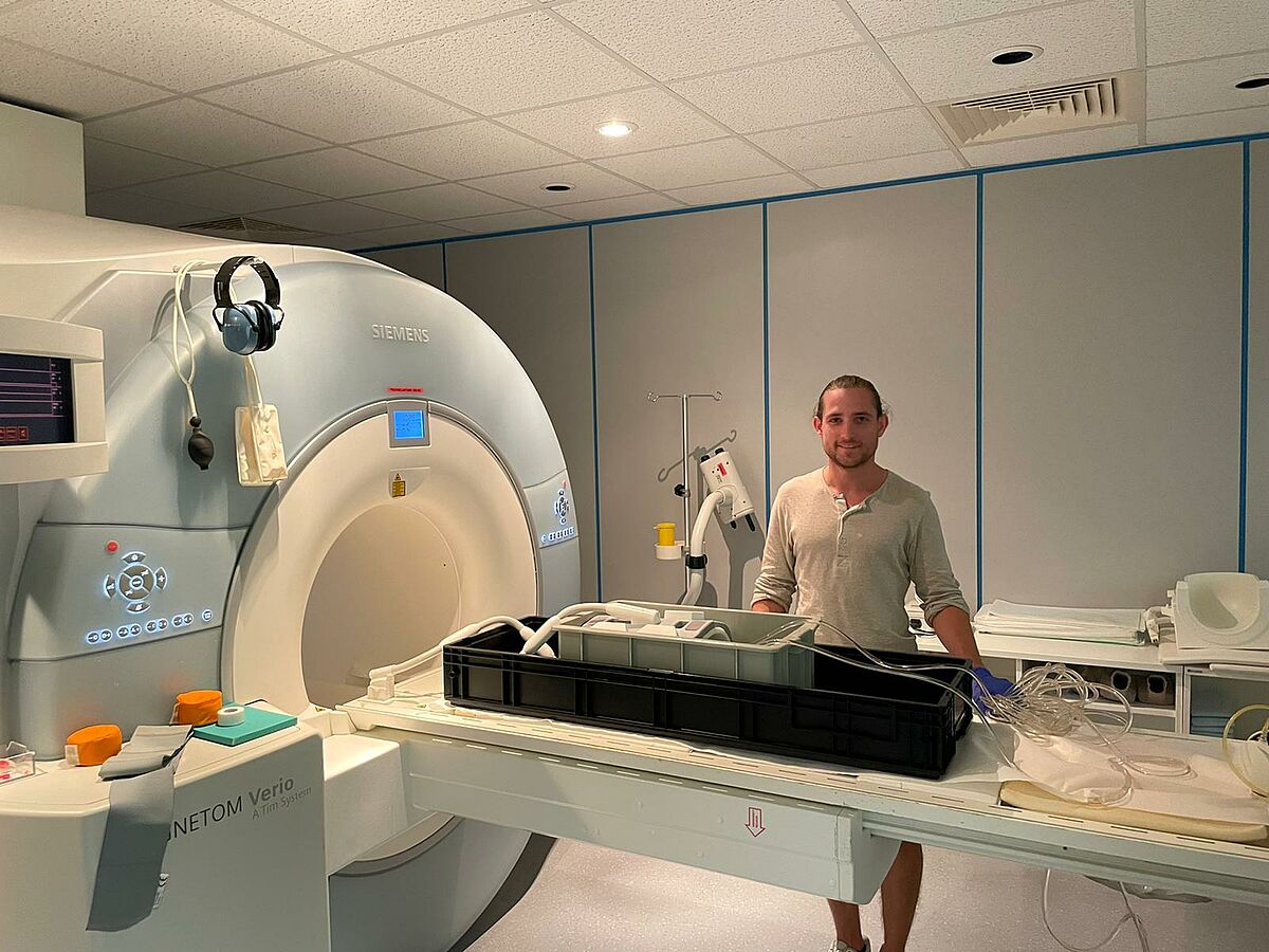
MRI (magnetic resonance imaging) is one of the medical imaging procedures that does not require X-rays. With an MRI, the entire body can be imaged, but individual areas of the body, such as the head or the knee, can also be depicted by means of the cross-sectional images.
Since the smallest building block of the human body are hydrogen atoms, it is possible to influence these with magnetic fields and radio waves by means of MRI. The small magnetic field created by the so-called spin can now be influenced with the help of a strong magnet. This magnet is located inside the MRI scanner - it encloses a tube that surounds the person being examined.
Due to the different composition of the individual organs, there is a different amount of hydrogen in it, which enables a good contrast between the organs during MRI. Soft tissues, such as the brain or internal organs, can be seen particularly well. Tumors and inflammations can also be depicted well in most cases, as their hydrogen content differs from that of healthy tissue.
The main research topics of our group are in the area of flow and diffusion imaging, as well as data processing of MRI data. More details are available here.
Contact
Prof. Dr. Susanne Schnell
Institute of Physics
Felix-Hausdorff-Str. 6
17489 Greifswald
phone: +49 3834 420 4740
fax: +49 3834 420 4701
susanne.schnelluni-greifswaldde
Study advisory:
Dr. Stephan König
phone: +49 3834 420 4706
studienberatung-medizinphysikuni-greifswaldde